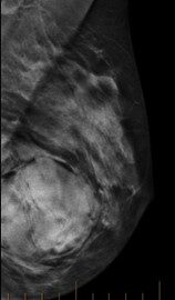
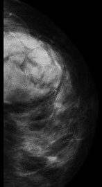
Patient presentation: Left breast lump on ultrasound for further investigation.
Requested examination: Bilateral mammogram
Examination Technique: Standard mammographic views with tomosynthesis
Findings:
Encapsulated left breast lesion correlating to the lesion identified in the previous ultrasound report. The density of this lesion is similar to the surrounding breast tissue. A differential diagnosis is a breast hamartoma.
A breast hamartoma is a benign, well-demarcated mostly encapsulated nodule appearing as a mass composed of haphazardly arranged breast tissue components.
A biopsy was arranged for tissue characterisation.
Biopsy result – overall features are that of a benign lesion. Most likely represents a hamartoma.
Breast tomosynthesis, also called three-dimensional (3D) mammography and digital breast tomosynthesis (DBT), is an advanced form of breast imaging, or mammography, that uses a low-dose x-ray system and computer reconstructions to create three-dimensional images of the breasts. Breast tomosynthesis aids in the early detection and diagnosis of breast disease. While mammography is the chosen screening tool for breast cancer available today, it does not detect all breast cancers. Breast tomosynthesis overcomes some of the limitations of standard mammography, but it is not yet available in all imaging facilities.
Synergy Radiology offers this service at our Campsie, Norwest and Rouse Hill locations.

