Case Studies


Patient presentation:

  • Suffering from a persistent cough after inhaling a lobster shell
  • Presented for a CT scan of the chest

A chest CT scan was performed without contrast, showing a foreign body in the right lower lobe bronchus, consistent with a piece of lobster shell. There was extensive mucous demonstrated throughout the right lower lobe. The patient was referred to emergency for further assessment.


Patient presentation:

  • Left sided lower neck pain, worsening with movement.
  • Lower back pain, radiating to both legs on walking and when standing for long periods of time.
  • Symptoms started after a workplace injury.

MRI Cervical and Lumbar spines:

  • Shows inflammation around the left C5/6 and left C6/7 facet joints, which is most likely the cause of the lower left neck pain.
  • Disc bulge at L4/5 level, causing compression of the exiting nerves to both sides, which is most likely the cause of the lower back and lower limb symptoms.

Requested cortisone injections:

  • CT guided left C5/6 and C6/7 facet joint injections – to treat left lower neck symptoms.
  • CT guided L4/5 level epidural injection – to treat lower back and bilateral leg symptoms.

The injections were performed on two separate days for the two different regions.

Cortisone is an anti-inflammatory to help treat joint and nerve injury/inflammation. It is administered via a needle, which is positioned by a radiologist under CT guidance. When the correct position has been obtained, a combination of anaesthetic and cortisone is injected which is designed to relieve the symptoms. This may take up to a week to reach full effect.

Our workers compensation cases are dealt with by dedicated staff who are specifically trained to assist in liaising with claims officers for pre-approval and to book an appointment and follow up with the referring doctor. All that is needed is a request from a medical practitioner, patient contact details and insurance details.

To make a workers compensation appointment, please contact our team:

Phone: (02) 8889 6317
Fax: (02) 8889 6333
Email: [email protected]


Patient presentation: Left breast lump on ultrasound for further investigation.

Requested examination: Bilateral mammogram

Examination Technique: Standard mammographic views with tomosynthesis

beast tomosynthesis

Findings:

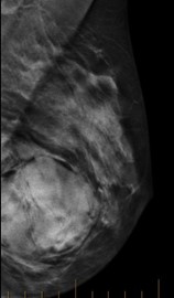
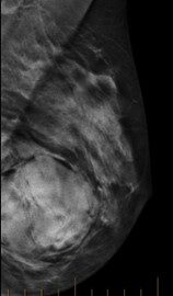
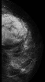
Encapsulated left breast lesion correlating to the lesion identified in the previous ultrasound report. The density of this lesion is similar to the surrounding breast tissue. A differential diagnosis is a breast hamartoma.

A breast hamartoma is a benign, well-demarcated mostly encapsulated nodule appearing as a mass composed of haphazardly arranged breast tissue components. 

A biopsy was arranged for tissue characterisation.

Biopsy result – overall features are that of a benign lesion. Most likely represents a hamartoma.

Breast tomosynthesis, also called three-dimensional (3D) mammography and digital breast tomosynthesis (DBT), is an advanced form of breast imaging, or mammography, that uses a low-dose x-ray system and computer reconstructions to create three-dimensional images of the breasts. Breast tomosynthesis aids in the early detection and diagnosis of breast disease. While mammography is the chosen screening tool for breast cancer available today, it does not detect all breast cancers. Breast tomosynthesis overcomes some of the limitations of standard mammography, but it is not yet available in all imaging facilities. 

Synergy Radiology offers this service at our Campsie, Norwest and Rouse Hill locations.